Key points
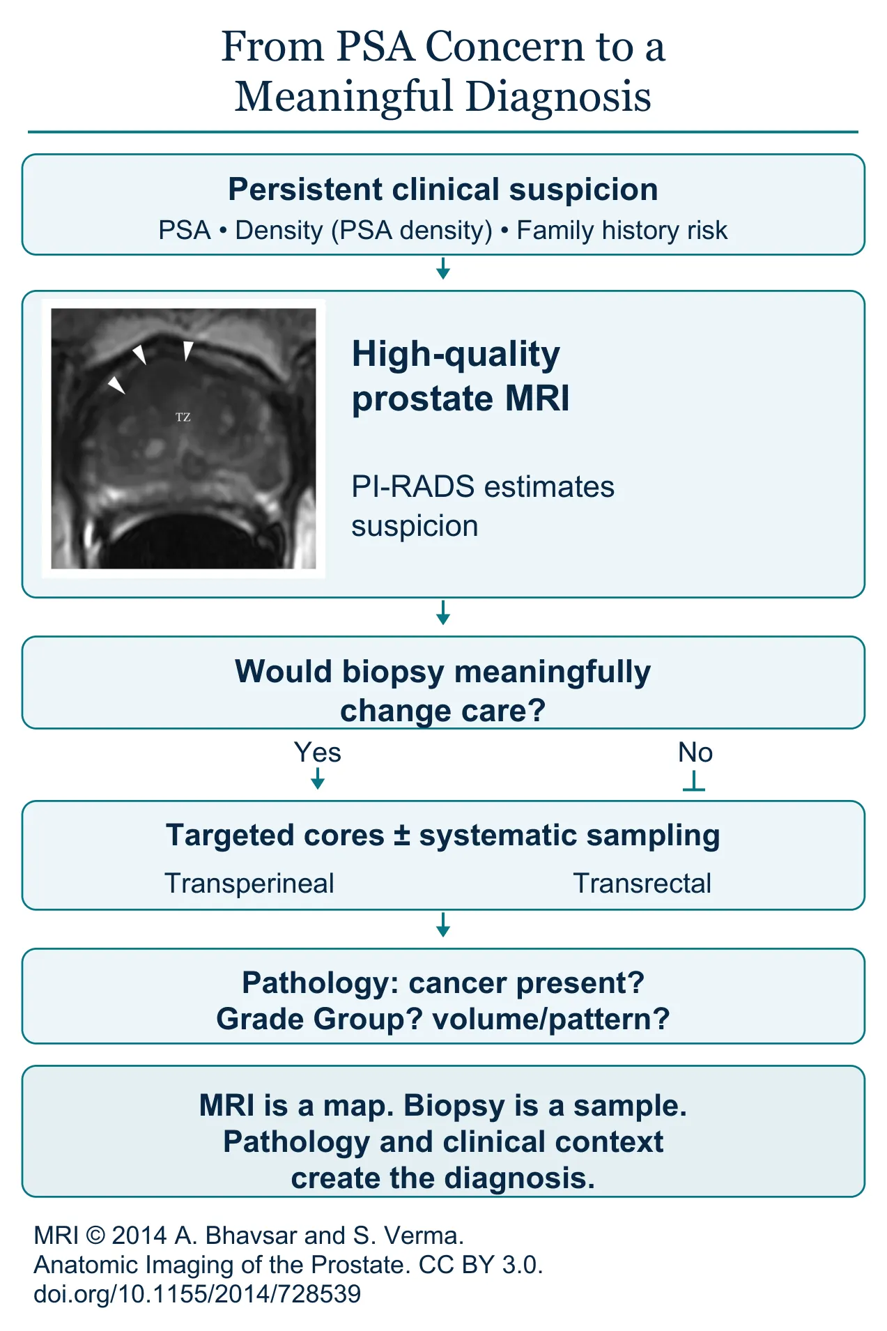
- A PI-RADS score estimates imaging suspicion; it is not a Gleason grade, Grade Group or stage.
- A highly suspicious lesion can prove benign, and a reassuring scan can miss cancer.
- Targeted cores sample the MRI lesion; systematic cores sample regions that may hide cancer not visible on MRI.
- Biopsy can be done through the rectum or the perineal skin; the route is a safety and access decision.
- Grade Group, the amount and distribution of cancer, PSA, MRI and stage together define risk.
- A negative MRI or biopsy lowers risk without always eliminating it.
From suspicion to tissue
MRI improves the map. Biopsy establishes the tissue diagnosis. Neither should be interpreted alone. It is common to read an MRI score as if it were a cancer stage and to believe the diagnosis is already complete. It is not. MRI estimates how suspicious an area appears and can help find where clinically significant cancer may be and guide sampling, but it does not provide the microscopic diagnosis. A highly suspicious lesion can prove benign. A reassuring scan can miss cancer. Imaging changes probability; tissue establishes what the cells actually are.
What PI-RADS means
Multiparametric prostate MRI evaluates anatomy and tissue behavior through several imaging sequences. Radiologists commonly summarize suspicion using PI-RADS, a structured scale from very low to very high likelihood that a clinically significant cancer is present. The score is not a Gleason grade, Grade Group or stage. Image quality and the reader’s expertise matter, as do prostate size, inflammation, prior biopsy and where the lesion lies. If the MRI is poor quality, no targeting system can rescue it.
Will a biopsy change your care?
Before biopsy, the clinical question should be explicit. Are we looking for cancer likely to affect how long or how well you live? Would you be willing and medically able to pursue surveillance or treatment if such cancer were found? When other illnesses make a tissue diagnosis unlikely to change care, more testing can create burden without benefit.
Targeted and systematic sampling
An MRI-targeted biopsy directs cores toward a suspicious lesion. Systematic sampling takes tissue from predefined regions that may harbor cancer not visible on MRI. Depending on whether this is a first or repeat biopsy, the MRI findings and your risk, the two approaches may be combined. More sampling can improve detection, but it also increases discomfort and bleeding and finds more small, slow-growing cancers that may never matter. The plan should be purposeful.
Choosing the biopsy route
Biopsy can be performed through the rectum or through the skin of the perineum, between the scrotum and the anus. Both routes can detect cancer. Transperineal techniques have become increasingly prominent because the needle avoids passing through rectal bacteria, and they may improve access to some front and lower regions of the gland. They need appropriate equipment, positioning, anesthesia and experience. Transrectal biopsy is still used in selected settings with infection-prevention protocols. The route should be discussed as a safety and access decision, not merely a habit.
Preparing for the procedure
Preparation should be tailored, not reduced to a generic instruction sheet. Tell the team about blood thinners, prior infections after biopsy, resistant organisms, medication allergies, urinary retention, cardiac devices, immune suppression and anesthesia concerns. Never stop an anticoagulant or antiplatelet drug without a coordinated plan. If a rectal route is planned, infection prevention deserves explicit attention; if a transperineal route is chosen, positioning, local anesthesia or sedation and temporary urinary effects should still be discussed.
Expect some temporary effects, including blood in the urine or semen, soreness and urinary symptoms. Fever, shaking chills, inability to urinate or significant deterioration after biopsy need prompt medical contact.
Arrange the result conversation before the procedure. Reading a pathology report through a portal without context can turn unfamiliar terms into unnecessary terror.
Reading the pathology report
The pathology report does more than say yes or no. Grade Group describes how abnormal and aggressive the sampled cancer appears. The amount and distribution of cancer, the number and location of positive cores, the percentage involvement, any higher-risk microscopic patterns, MRI findings, PSA, clinical stage and sometimes genomic or genetic information all contribute to risk classification. These are parts of a risk model, not isolated verdicts. A biopsy samples portions of the prostate; it does not examine the entire gland, so apparent precision must be balanced with sampling uncertainty.
A negative biopsy can be reassuring without ending follow-up forever. If PSA density, MRI, inherited risk or another finding remains concerning, follow-up may involve repeat PSA, repeat imaging, biomarkers, pathology review or a selected repeat biopsy. On the other hand, automatic repeat biopsy based on PSA alone can expose you to repeated harm without adding enough information.
When the map and the sample disagree
Disagreement between imaging and pathology is common enough to deserve a plan. A highly suspicious lesion with benign targeted cores may reflect inflammation, a targeting error, sampling limits or a false-positive image. A low-grade biopsy in a lesion that looks aggressive may need expert radiology and pathology review or more workup. Significant cancer found outside an MRI target is a reminder of why systematic sampling may still matter in selected settings.
Disagreement does not mean repeating every test. The better question is which component is least secure. Was the MRI high quality and read by an experienced radiologist? Was the lesion actually sampled? Was pathology reviewed by someone with genitourinary expertise? Does PSA density still support concern? The next step should repair the weakest link rather than restart the entire process.
A diagnosis is complete only when the pathology has been connected back to you. A small amount of low-grade cancer creates a different future from extensive high-grade disease. A positive result begins the treatment discussion, but the distribution and biology determine its direction. The goal of modern diagnosis is not simply to find more prostate cancer, but to find the cancer that matters while reducing unnecessary biopsy and overdiagnosis.
Questions to ask your urologist
- What does my MRI show, what is its PI-RADS score, and what might it miss?
- Would a biopsy change my care?
- Why does the proposed biopsy route fit my situation, and how will infection and retention risk be reduced?
- Will you take targeted cores, systematic cores or both?
- Who will explain the complete pathology report, and when?
- If the MRI and biopsy disagree, which part should be reviewed?