Key points
- The disappearance of blood tells you active bleeding stopped, not why it started.
- Painless bleeding is not necessarily reassuring; some conditions worth excluding cause no discomfort.
- A dipstick is a reason to look more carefully; microscopic examination confirms red blood cells.
- Blood thinners can reveal bleeding but do not explain its source, and should never be stopped on your own.
- Cystoscopy and upper-tract imaging answer complementary questions; one does not replace the other.
- Reassurance is most valuable when it follows an appropriate evaluation rather than replacing one.
It happened once, and then it stopped
Many people see red urine one morning and find it clear again by lunchtime. It is tempting to cancel the appointment. But bleeding can be intermittent. A stone can move, inflamed tissue can settle, a prostate vessel can stop leaking, and a bladder or kidney tumor can bleed one day and stay silent for weeks. The disappearance of blood tells you that active bleeding stopped. It does not tell you why it started.
The absence of pain is not necessarily reassuring either. Infection and stones often hurt; some of the conditions most worth excluding cause no discomfort at all. This does not mean blood in the urine usually means cancer. It does not. Infection, stones, prostate bleeding, kidney disease, trauma, exercise and procedures are common causes. The point is neither to panic nor to invent reassurance before the source has been considered.
First, make sure it is blood
Red or brown urine is not always caused by red blood cells. Certain foods, pigments and medications change urine color. Menstrual or vaginal bleeding can contaminate a sample, rectal bleeding can be mistaken for urinary bleeding, and very concentrated urine can look dark. A dipstick detects a chemical signature of blood but does not always confirm intact red cells; microscopic examination does. A positive dipstick is a reason to look more carefully, not a diagnosis.
Visible and microscopic blood
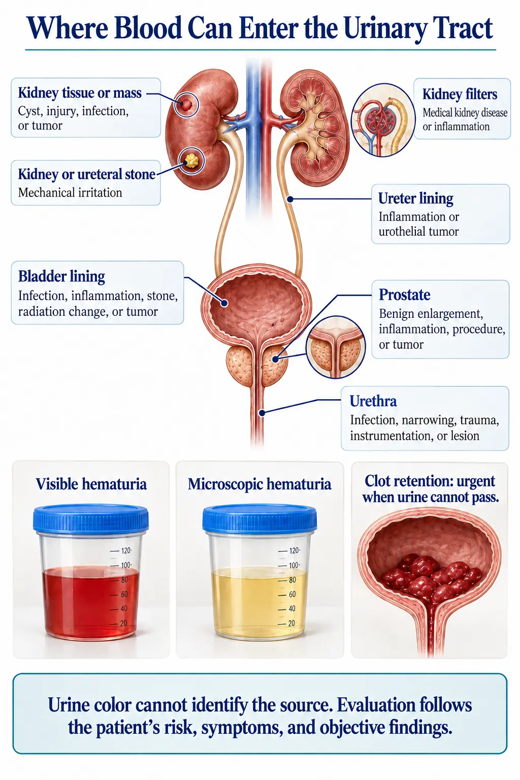
Visible hematuria turns urine pink, red, rust, tea-colored or brown, sometimes with clots. Even one episode deserves medical attention when no clear temporary cause has been confirmed.
Microscopic hematuria cannot be seen; it is found when a properly collected sample shows red cells under the microscope. How it is handled depends on the amount and persistence of blood and on your risk: age, smoking history, prior visible bleeding, pelvic radiation, certain chemical or medication exposures, family history and urinary symptoms. Risk-based evaluation protects low-risk people from unnecessary testing while directing a fuller workup to those most likely to benefit.
The pattern around the blood
Blood with severe flank pain that comes in waves and travels toward the groin makes a stone more likely. Burning, urgency and frequency suggest infection or inflammation. Fever, chills, vomiting or confusion can signal a more serious infection, especially if drainage is blocked. Blood after trauma, a catheter, surgery or strenuous exercise has its own explanations, but persistent, heavy or recurring blood still deserves another look. Painless visible blood is exactly the pattern that leads a urologist to examine the bladder lining and upper urinary tract carefully.
Common sources
- Infection can cause bleeding. After treatment, a repeat microscopic urinalysis confirms the blood is gone; if it persists, the first explanation was incomplete.
- Stones can irritate the kidney, ureter or bladder, with or without severe pain.
- The prostate, when enlarged or inflamed, may have fragile vessels; a recent biopsy, catheter or procedure can cause temporary blood.
- Exercise, menstruation, trauma and prior radiation are other possibilities.
- The kidney filters themselves can leak blood. Protein in the urine, abnormal kidney function, high blood pressure or swelling may point to medical kidney disease and a nephrologist’s involvement.
Blood thinners reveal bleeding; they do not explain it
Anticoagulant and antiplatelet medicines can make bleeding more likely or more visible, and may reveal a lesion that would otherwise have bled less. They do not protect you from stones, tumors, infection or prostate or kidney disease, so you deserve the same evaluation as anyone else. Never stop a blood thinner on your own; doing so can risk stroke, clots or heart attack. Your prescriber and urologist should weigh that decision together.
Looking above and below
No single test examines the whole urinary tract equally well. Cystoscopy looks directly at the urethra, prostate channel and bladder lining. Upper-tract imaging looks at the kidneys and ureters; it may be ultrasound for selected intermediate-risk microscopic blood, or CT urography for higher-risk evaluation and visible blood, with MR urography when CT contrast or radiation is unsuitable. A normal CT does not always replace cystoscopy, and a normal cystoscopy does not examine the kidneys.
For confirmed microscopic blood without a temporary explanation, low-risk patients may simply have repeat urine testing, intermediate-risk patients are usually offered cystoscopy and ultrasound, and high-risk patients generally need cystoscopy and more detailed imaging. In selected intermediate-risk situations, validated urine tests can inform a shared decision, but they are not universal substitutes for looking.
If cystoscopy finds a suspicious bladder lesion, it is usually removed or sampled in a procedure called TURBT so a pathologist can report both its grade (how aggressive the cells look) and its stage (how deeply it has grown).
When the evaluation finds nothing dangerous
A complete evaluation does not always find one cause. Learning that the bladder lining is normal, the kidneys show no concerning mass and kidney function is stable still changes your risk substantially. A new episode of visible blood, more microscopic blood or new symptoms can justify a fresh look later. Treatment follows the source: infection as infection, stones by size and location, prostate bleeding at the prostate, kidney-filter disease through a medical kidney pathway, and tumors through their own staging and treatment.
Questions to ask your urologist
- Was the blood confirmed under the microscope or only by dipstick?
- Is my bleeding visible or microscopic, and how do my risk factors shape the next step?
- Do I need cystoscopy, upper-tract imaging, repeat urine testing or a combination?
- Could this be coming from the kidney filters, and should a nephrologist be involved?
- If infection is treated, when should my urine be rechecked?
- What should I do if I pass clots or cannot urinate?