Key points
- Imaging is not a ladder with MRI always above CT and CT above ultrasound.
- Ultrasound avoids radiation and is useful for kidneys, bladder, prostate and scrotum, but may miss small ureteral stones.
- CT is rapid and highly informative for stones, trauma and masses; the protocol determines what it can answer.
- MRI gives detailed soft-tissue information without radiation, but takes longer and needs review of implants and gadolinium use.
- Contrast is neither harmless nor always dangerous; the decision should be individualized.
- Bring the actual images, not only reports, and know who owns follow-up of incidental findings.
Choose the test that can see the question
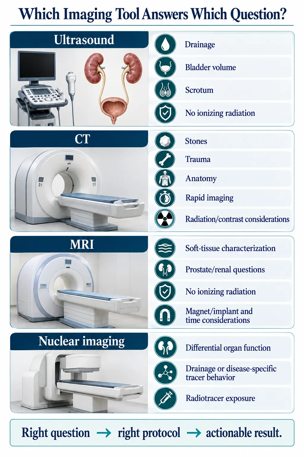
The most detailed scan is not always the best scan. Imaging is not a ladder on which MRI always sits above CT and CT above ultrasound. Each study answers a different anatomical or functional question, and the best test is the least burdensome one that can change the next decision. Ask for the imaging question in one sentence; the answer should explain why a particular test and protocol fit it.
What each method offers
- Ultrasound uses sound waves and avoids ionizing radiation. It can assess the kidneys, swelling from blocked drainage, bladder volume, urine left after voiding, prostate volume, blood flow in the scrotum and selected masses. But it may miss small stones in the ureter or fail to fully characterize complex tissue.
- CT is rapid and highly informative for stones, trauma, bleeding, masses and anatomy. Radiation and iodinated contrast are real considerations, but avoiding CT can also carry risk when it is the test most likely to find a dangerous condition.
- MRI provides detailed soft-tissue information without ionizing radiation and is central to selected prostate, kidney, pelvic and neurologic questions. It takes longer and requires review of implants, metal, claustrophobia, and gadolinium contrast use in kidney disease or pregnancy.
- Nuclear studies use small amounts of radioactive tracer to assess how much each kidney contributes to function, how well the kidneys drain, or disease-specific biology.
Protocol is part of the prescription
Your clinician should specify the question and the protocol rather than simply ordering “a scan.” “CT scan” and “MRI” are incomplete orders. A noncontrast CT can be excellent for finding stones yet inadequate for characterizing how a kidney mass enhances with contrast. A hematuria protocol evaluates the urinary tract differently from a routine abdominal scan. Prostate MRI needs appropriate sequences, equipment and interpretation. Nuclear drainage testing depends on technique, hydration, medications and the question being asked. The wrong CT phase or an MRI without the needed sequences can create an expensive test that never answers the clinical problem.
That is why carrying a prior report is sometimes not enough. The actual images allow a radiologist or specialist to see whether the needed anatomy was captured, whether a finding truly changed and whether repeating the study is necessary. Bring prior images whenever you can, especially when a finding is being compared over time, in a viewable electronic format, along with the reports and the dates of each study.
Contrast decisions should be individualized
Contrast is neither harmless nor always dangerous. The decision considers your kidney function, any prior reactions, the contrast agent, the imaging question, pregnancy, medications and whether a noncontrast alternative can provide equivalent information. Modern risk assessment distinguishes people with stable kidney function from those with acute kidney injury or severely limited reserve.
Avoiding indicated contrast can produce an unreadable or incomplete study and delay diagnosis. Giving it without reviewing relevant risks can cause preventable harm. The right approach is to define the benefit, assess you as an individual, use an appropriate protocol and apply current precautions, rather than rely on a blanket rule. Tell the team about kidney disease, prior contrast reactions, implants, pregnancy and claustrophobia in advance.
Incidental findings need their own triage
Incidental findings are common. An unexpected cyst, nodule, calcification or anatomical variation may need no action, a repeat scan at an interval, a different type of imaging or specialist review. The radiologist’s recommendation is a starting point, not an independent diagnosis. Follow-up should reflect the finding’s actual risk, the quality of the original study, prior imaging, your age and other illnesses, and whether more information would change care.
The report should be connected back to the reason the scan was done, so that unrelated abnormalities are neither ignored nor allowed to take over your care. Leave knowing who owns any follow-up. “Correlate clinically” is not a plan until a clinician connects the image to you.
Balancing the risks
Imaging methods provide complementary information, and their usefulness depends partly on protocol and contrast selection. The decision should weigh radiation and contrast risks against the consequences of missing disease. Some questions cannot be answered from outside the body at all; then office testing, such as cystoscopy or urodynamics, may be the next step, but it too should follow a defined question.
Questions to ask your urologist
- What question is this scan meant to answer?
- Why is this type of imaging and this protocol the right fit?
- How do radiation, contrast, implants, kidney function or pregnancy affect the choice?
- Do you need my prior images, not just the reports?
- Who will follow up any incidental finding, and when?