Key points
- Fever with an obstructed urinary system is an emergency.
- Pain relief does not always mean the kidney is draining; follow-up imaging or documented passage matters.
- Forcing excessive water does not push an obstructing stone through and may worsen pain.
- In an infected, blocked kidney, the first procedure may drain the kidney without removing the stone.
- Normal dietary calcium, taken with food, often helps; blanket calcium restriction can be counterproductive.
- Prevention is a feedback loop: measure, intervene, remeasure and adjust.
Pain is not the only measure of danger
Stone pain usually begins when a stone blocks the ureter, the tube from the kidney to the bladder, and pressure builds above it. The pain may move from the back or side toward the lower abdomen, groin, testicle or labia as the stone travels. Nausea, vomiting, urgency, blood in the urine and restlessness are common, though no symptom pattern proves a stone is present.
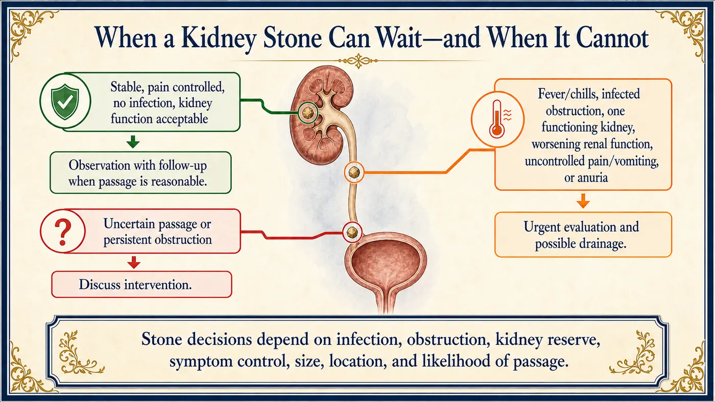
A stone becomes urgent when it threatens drainage, kidney function, infection control, hydration or your ability to stay stable, not simply when it hurts. A comfortable patient can still have a persistent blockage, and a patient in dramatic pain may have a stone likely to pass.
Finding the stone
Imaging defines size, location, blockage and other possible diagnoses. CT is highly sensitive. Ultrasound avoids radiation and is especially valuable in pregnancy and some follow-up settings, though it may miss stones in the ureter. Urinalysis, culture, kidney function, a pregnancy test and blood count are chosen according to how you present.
When a stone can wait
Observation may be appropriate when pain is controlled, there is no infection, kidney function is safe and passage is plausible. Treatment may include pain and nausea control, enough fluid to avoid dehydration and, in selected cases, a medication to help passage. Forcing excessive water does not push a blocking stone through and may worsen pain.
Watching a stone is an active plan, not “come back if it hurts.” You should know how pain will be managed, how to catch the stone if possible, when follow-up happens and how passage or relief of blockage will be confirmed, because a blockage can occasionally persist after pain improves. People with one functioning kidney, reduced kidney function, pregnancy, recurrent infection or unreliable access to emergency care need a lower threshold for reassessment.
When it cannot wait
Infection behind a blockage is an emergency. The immediate job may be drainage with a ureteral stent (a thin internal tube) or a nephrostomy (a tube through the back), not stone removal. The stone may be treated later, once infection and the body have settled; combining removal with an unstable infection can increase risk. Stents can cause frequency, urgency, blood, flank discomfort during urination and poor sleep. Knowing that helps you tell expected recovery from a new emergency, but fever, inability to urinate, uncontrolled pain or getting worse still need prompt contact.
Ureteroscopy, shock-wave lithotripsy and percutaneous stone removal suit different stones and anatomies. The choice depends on size, location, density, anatomy, bleeding risk, infection, pregnancy, kidney reserve and your priorities.
Preventing the next stone
A stone is a crystallized metabolic history. It forms when urine becomes concentrated and chemically favorable for crystals. Low urine volume is common, but calcium, oxalate, uric acid, citrate, sodium, acidity, infection, anatomy, bowel disease, medications and inherited conditions also affect risk. “Drink more water” is useful advice, but not a complete plan.
- Keep the stone when you can; analysis tells you what you made.
- Blood tests may reveal kidney, calcium or uric-acid problems.
- A 24-hour urine collection is especially helpful for recurrent stones, high-risk first stones, a single kidney or unusual stone types. It should reflect a typical day; a quiet Sunday may not represent a hot workday.
Making prevention specific
Fluid should produce consistently dilute urine across workdays, exercise, heat and sleep, not just meet a generic bottle count. Sodium from restaurant food, packaged food and sauces can raise urinary calcium. Normal dietary calcium, taken with food, often helps bind oxalate in the gut; indiscriminate calcium restriction may backfire. Oxalate advice should target your meaningful sources rather than turn a long food list into fear. Excess animal protein, a high uric-acid load, low fruit and vegetable intake, bowel disease and certain supplements may matter depending on your stone type.
Medication is chosen for a defined abnormality or recurrence pattern, such as thiazide-type therapy, citrate or alkali, or uric-acid–lowering treatment, and it needs monitoring. A recent major trial (NOSTONE) did not show a substantial reduction in recurrence with hydrochlorothiazide compared with placebo in recurrent calcium-stone formers, and some side effects were more common. That makes careful selection and follow-up especially important, though it does not show that every thiazide regimen is ineffective.
Follow-up closes the loop. A repeat urine collection shows whether treatment changed the intended chemistry. The same advice cannot work equally for a low-volume calcium oxalate former, a uric-acid former with acidic urine and someone with infection-related stones.
Questions to ask your urologist
- Does infection, blockage or threatened kidney function change how urgent my stone is?
- How likely is my stone to pass, and how will we confirm that it has?
- How do ureteroscopy, shock-wave lithotripsy and percutaneous removal compare for my stone?
- What type of stone did I form, and would a 24-hour urine test change my prevention plan?
- Which abnormality is my plan targeting, and when will we remeasure?